
Skip the wait times and get your X-Ray as soon as you need it. X-ray services are available Monday to Friday at our Calgary South Clinic. Saturday, limited coverage, call for availability. We accept all X-ray requisitions with appointments or walk-ins.
Please note: Available in Calgary clinic only.
Healthcare providers? Download a requisition →
Depending on the nature of your X-Ray, you may be required to follow exam preparation instructions. Click on your exam below to find out how to prepare.
We provide a wide range of x-ray scans to detect fractures, organ enlargement or displacement, foreign objects, gas patterns indicative of bowel conditions, calcifications, and soft tissue abnormalities in the abdominal region.
Abdomen x-rays are non-invasive imaging tests that use X-ray radiation to capture static images the abdominal area, including organs like the liver, kidneys, and intestines. They help diagnose conditions such as bowel obstructions, abdominal masses, and fractures.
This painless procedure is safe and commonly used for assessing abdominal health.A Chest PA (Posteroanterior) / Lateral X-Ray is a common diagnostic procedure used to capture images of the chest area, including the heart, lungs, ribs, and diaphragm.
In a PA X-ray, the X-ray beam passes from the back to the front of the chest, while in a lateral X-ray, the beam passes from one side of the body to the other.
Doctors might recommend a Chest PA / Lateral X-Ray to evaluate various conditions such as:
Respiratory issues: To assess lung health and detect conditions like pneumonia, tuberculosis, or chronic obstructive pulmonary disease (COPD).
Cardiac abnormalities: To examine the heart’s size, shape, and position within the chest cavity, and to detect signs of heart failure or structural abnormalities.
Trauma or injury: To identify fractures, dislocations, or other injuries to the ribs, sternum, or other chest structures.
Monitoring: To monitor the progression of certain diseases, such as lung cancer or heart disease, or to assess the effectiveness of treatment.
A Kidney, Ureter & Bladder (KUB) X-Ray is a diagnostic imaging procedure used to assess the structures of the urinary system, including the kidneys, ureters, and bladder.
During the KUB X-Ray, the patient lies on an X-ray table while images are taken from the front to capture the abdomen.
Doctors might recommend a KUB X-Ray for several reasons:
Kidney stones: To detect the presence of kidney stones and assess their size, location, and number within the kidneys or along the urinary tract.
Urinary tract infections (UTIs): To identify signs of urinary tract infections, such as urinary obstruction or inflammation, which may require further evaluation and treatment.
Renal abnormalities: To evaluate the size, shape, and position of the kidneys and detect any abnormalities such as cysts, tumors, or anatomical variations.
Urinary system injuries: To diagnose injuries to the kidneys, ureters, or bladder resulting from trauma or accidents.
Monitoring: To monitor the progression of certain conditions affecting the urinary system, such as kidney stones or kidney function disorders.
A Sternum X-Ray examines the sternum, also known as the breastbone, which is located at the center of the chest.
During the X-ray, the patient typically stands upright or lies on an X-ray table, and images are taken from the front and side to capture the entire sternum.
Doctors might recommend a Sternum X-Ray for several reasons:
Chest pain or trauma
Respiratory issues
Bone abnormalities
Monitoring
A SC (sternoclavicular) Joints X-Ray examines the sternoclavicular joints, which connect the sternum (breastbone) to the clavicles (collarbones).
During the X-ray, the patient typically stands upright or lies on an X-ray table, and images are taken from different angles to capture the sternoclavicular joints.
A doctor might recommend this X-Ray to assess joint pain, swelling, or limited motion in the shoulders or chest, to evaluate trauma-related injuries like fractures or dislocations, to identify signs of infection, or to detect abnormalities such as tumors or cysts around the sternoclavicular joints.
Skull X-rays are imaging tests used to detect fractures, bone abnormalities, sinus infections, dental issues, tumors, and foreign objects within the skull and surrounding areas.
A skull X-Ray captures images of the bones of the skull, including the cranium and facial bones.
During the X-ray, the patient typically sits or lies on an X-ray table, and images are taken from various angles to obtain a comprehensive view of the skull.
A doctor might recommend a skull X-Ray to evaluate head trauma for fractures or injuries, assess sinus problems or dental issues, and monitor conditions like bone tumors or developmental abnormalities affecting the skull bones.
A sinus X-Ray visualizes the sinus cavities, which are air-filled spaces in the skull located around the nose and eyes. The procedure involves capturing X-ray images of the frontal, maxillary, ethmoid, and sphenoid sinuses.
A doctor might recommend a sinus X-Ray to assess symptoms such as facial pain, congestion, or headaches, to evaluate sinus infections, inflammation, or abnormalities, and to guide treatment decisions such as antibiotics, sinus surgery, or sinus drainage procedures.
A Soft Tissue (Neck & Adenoids) X-Ray examines the soft tissues in the neck area, including the adenoids, which are glands located at the back of the throat.
During the X-ray, the patient typically stands or sits upright, and images are taken from various angles to capture the soft tissue structures.
A doctor might recommend this X-Ray to assess symptoms such as difficulty swallowing, breathing problems, or recurrent throat infections. It helps evaluate adenoid enlargement, infections, or blockages in the airways and guides treatment decisions.
A nasal bone X-Ray visualizes the nasal bones, which form the bridge and structure of the nose.
During the X-ray, the patient typically sits or stands with their head positioned to capture images of the nasal region.
A doctor might recommend a nasal bone X-Ray to evaluate nasal fractures resulting from trauma or injury to the nose. It helps assess the alignment, shape, and integrity of the nasal bones, aiding in treatment decisions such as realignment or surgical intervention.
A mandible X-Ray examines the mandible, or lower jawbone. During the X-ray, the patient typically sits or stands while images are taken from various angles to capture the structure of the mandible.
A doctor might recommend a mandible X-Ray to assess trauma or injury to the lower jawbone, such as fractures or dislocations resulting from accidents, falls, or sports injuries. It helps identify the extent and location of the injury, guiding treatment decisions such as realignment, surgical intervention, or immobilization.
A Temporomandibular (TM) Joint X-Ray is a diagnostic imaging procedure used to visualize the temporomandibular joints, which connect the jawbone to the skull. During the X-ray, the patient typically sits or stands while images are taken from various angles to capture the TM joint.
A doctor might recommend a TM Joint X-Ray to assess symptoms such as jaw pain, clicking or popping sounds, or difficulty chewing or opening the mouth. It helps evaluate the TM joint for signs of arthritis, inflammation, dislocation, or structural abnormalities, guiding treatment decisions such as physical therapy, medications, or surgery.
An Orbits X-Ray examines the eye sockets or orbits. During the X-ray, the patient typically sits or stands while images are taken from various angles to capture the structures surrounding the eyes.
A doctor might recommend this to evaluate symptoms such as eye pain, swelling, or changes in vision. It helps assess for fractures, tumors, or other abnormalities affecting the bones or soft tissues surrounding the eyes, guiding treatment decisions such as surgical intervention or further imaging studies.
A Mastoids X-Ray examines the mastoid bones, which are located behind the ears. During the X-ray, the patient typically sits or stands while images are taken to capture the mastoid region.
A doctor might recommend a Mastoids X-Ray to assess symptoms such as ear pain, swelling, or discharge. It helps evaluate for infections, fractures, tumors, or other abnormalities affecting the mastoid bones or surrounding structures, guiding treatment decisions such as antibiotic therapy, surgical intervention, or further imaging studies.
A Spine & Pelvis X-Ray is a diagnostic imaging procedure used to examine the bones and alignment of the spine and pelvis. It helps evaluate symptoms such as back pain, stiffness, or limited mobility.
A cervical spine X-ray assesses the bones and alignment of the cervical spine, which is the portion of the spine located in the neck. During the X-ray, the patient typically sits or stands while images are taken from various angles to capture the cervical vertebrae.
A doctor might recommend a cervical spine X-ray to evaluate symptoms such as neck pain, stiffness, or numbness and tingling in the arms or hands.
A thoracic spine X-ray is used to assess the bones and alignment of the thoracic spine, which is the portion of the spine located in the mid-back. During the X-ray, the patient typically stands or sits while images are taken from various angles to capture the thoracic vertebrae.
This exam helps evaluate symptoms such as mid-back pain, stiffness, or difficulty breathing.
A Lumbar Spine X-ray assesses the five vertebrae of the lower back (L1 through L5) to evaluate bone structure and alignment. During the procedure, the patient typically lies on an X-ray table while images are captured from multiple angles to provide a clear view of the lumbar column.
It is important to note that a Lumbar Spine X-ray does not automatically include the sacrum or coccyx. By ordering a Lumbar Spine, you will get a Lumbar spine. If the Sacrum and Coccyx are required, then Dr’s will need to order this to be clear of what they need.
A Sacrum/Coccyx X-Ray assesses the bones and alignment of the sacrum and coccyx, which are the lowest segments of the spine located at the base of the vertebral column. During the X-ray, the patient typically lies on an X-ray table while images are taken to capture the sacrum and coccyx.
This type of X-ray helps evaluate symptoms such as tailbone pain, lower back pain, or discomfort in the pelvic region.
A SI (sacroiliac) Joints X-Ray assesses the sacroiliac joints, which connect the sacrum to the pelvis. During the X-ray, the patient typically lies on an X-ray table while images are taken to capture the sacroiliac joints.
This type of X-ray helps evaluate symptoms such as lower back pain, buttock pain, or hip pain.
An AP (anteroposterior) Pelvis X-Ray visualizes the pelvis from the front to the back. During the X-ray, the patient typically lies on an X-ray table while images are taken to capture the entire pelvis.
This type of X-ray helps evaluate symptoms such as hip pain, groin pain, or difficulty walking. It aids in assessing for conditions such as fractures, dislocations, or abnormalities in the pelvis, hip joints, or surrounding structures.
A Pelvis & Hip X-Ray examines the bones and joints of the pelvis and hips. During the X-ray, the patient typically lies on an X-ray table while images are taken to capture the entire pelvis and hip joints.
This type of X-ray helps evaluate symptoms such as:
hip pain
groin pain
difficulty walking, or limited range of motion in the hips
fractures
dislocations
arthritis, or other abnormalities affecting the pelvis, hip joints, or surrounding structures.
A Scoliosis Series X-Ray is a diagnostic imaging procedure used to assess the curvature and alignment of the spine, particularly focusing on scoliosis, which is an abnormal sideways curvature of the spine. During the X-ray, the patient typically stands while images are taken to capture the entire spine from different angles.
This type of X-ray helps evaluate symptoms such as:
Back pain
Uneven shoulders or hips
Asymmetrical appearance of the spine
We provide a wide range of x-ray scans to detect fractures, organ enlargement or displacement, foreign objects, gas patterns indicative of bowel conditions, calcifications, and soft tissue abnormalities in the abdominal region.
A Shoulder X-Ray is a diagnostic imaging procedure used to assess the bones and joints of the shoulder. During the X-ray, the patient typically stands or sits while images are taken to capture various views of the shoulder joint.
It aids in identifying fractures, dislocations, arthritis, or other abnormalities affecting the bones, joints, or soft tissues of the shoulder. Symptoms evaluated include:
Shoulder pain
Swelling
Stiffness
Limited range of motion
A Clavicle X-Ray is a diagnostic imaging procedure used to assess the collarbone, or clavicle. During the X-ray, the patient typically stands or sits while images are taken to capture various views of the clavicle.
This type of X-ray helps evaluate symptoms such as pain, swelling, or deformity in the collarbone area. It aids in identifying fractures, dislocations, or other abnormalities affecting the clavicle.
Symptoms evaluated include:
Collarbone pain
Swelling around the collarbone
Deformity in the collarbone area
An AC (acromioclavicular) joints X-ray is a diagnostic imaging procedure used to assess the acromioclavicular joints, which connect the shoulder blade to the collarbone. AC joint X-rays are always performed on both sides (in pairs) for comparison.
During the X-ray, the patient typically stands while images are taken in both non-weight-bearing and weight-bearing positions. These comparative views help the radiologist determine if there is any joint separation or abnormality.
This type of X-ray helps evaluate symptoms such as shoulder pain, swelling, or tenderness localized to the AC joint area. It aids in identifying injuries such as sprains, separations, or fractures affecting the AC joint.
Symptoms evaluated include:
Shoulder pain localized to the AC joint
Swelling around the AC joint
Tenderness over the AC joint area
A Scapula X-Ray is a diagnostic imaging procedure used to assess the scapula, or shoulder blade. During the X-ray, the patient typically stands or sits while images are taken to capture various views of the scapula.
This type of X-ray helps evaluate symptoms such as shoulder pain, swelling, or tenderness localized to the scapular region. It aids in identifying fractures, dislocations, or other abnormalities affecting the scapula.
Symptoms evaluated include:
Shoulder pain localized to the scapular region
Swelling around the scapula
Tenderness over the scapula area
Elbow X-ray is a diagnostic imaging procedure used to assess the bones and joints of the elbow. The X-ray images are taken while the patient sits or stands, capturing various angles to visualize the elbow joint.
The exam identifies fractures, dislocations, arthritis, or other abnormalities affecting the elbow bones or joints. Symptoms evaluated include:
Elbow pain
Swelling
Tenderness
Limited range of motion
A forearm X-ray is a diagnostic imaging procedure used to evaluate the bones of the forearm. During the X-ray, the patient’s arm is typically positioned on an X-ray table, and images are taken to capture the forearm.
Forearm X-ray helps in assessing various conditions such as fractures, dislocations, or deformities affecting the bones of the forearm. Symptoms evaluated include:
forearm pain
swelling
tenderness
difficulty moving the arm.
A Wrist X-Ray is a diagnostic imaging procedure utilized to evaluate the bones and surrounding structures of the wrist.
During the X-ray, the patient typically positions their hand and wrist on a specialized plate, allowing images to be taken from multiple angles.
Symptoms evaluated include:
Wrist pain
Swelling around the wrist joint
Restricted movement of the wrist
Tenderness upon touching specific areas of the wrist
A Scaphoid X-ray assesses potential fractures or injuries to the scaphoid bone, which is located in the wrist.
Symptoms evaluated include:
Pain and tenderness in the wrist
Swelling around the wrist
Difficulty moving the wrist or thumb
Bruising or discoloration around the wrist area
Pain that worsens with gripping or bearing weight on the affected hand
A Hand X-Ray is a diagnostic imaging procedure used to evaluate the bones and structures of the hand and fingers.
During the X-ray, the patient will be sitting on a stool, with their hand on a specialized plate.
Symptoms evaluated include:
Pain, tenderness, or swelling in the hand or fingers
Difficulty moving the hand or fingers
Suspected bone abnormalities or growths
Evaluation of bone age in pediatric patients
A digits X-ray is a radiographic imaging procedure used to examine the bones, joints, and soft tissues of the fingers or toes, collectively known as digits. It is employed to diagnose and assess various conditions affecting these structures, including fractures, dislocations, arthritis, and abnormalities.
Symptoms evaluated include:
Pain, tenderness, or swelling in the fingers or toes
Difficulty moving the digits
Deformity or misalignment of the fingers or toes
Signs of trauma or injury, such as bruising or lacerations
Suspected bone abnormalities, growths, or foreign bodies
A lower extremities X-ray evaluates fractures, dislocations, arthritis, and bone abnormalities in the bones, joints, and soft tissues of the lower parts of the body, including the hips, pelvis, thighs, knees, lower legs, ankles, & feet.
A hip X-ray assesses the bones, joints, and surrounding tissues of the hip. It helps healthcare providers diagnose and evaluate various conditions affecting the hip joint, including fractures, dislocations, arthritis, and bone abnormalities.
A femur X-ray is a radiographic imaging procedure used to examine the femur, which is the large bone located in the thigh. It helps healthcare providers diagnose and evaluate various conditions affecting the femur, including fractures, tumors, infections, and bone abnormalities.
A femur X-ray is used to examine the femur, which is the large bone located in the thigh. It helps healthcare providers diagnose and evaluate various conditions affecting the femur, including fractures, tumors, infections, and bone abnormalities.
A Tibia and Fibula X-ray is a radiographic imaging procedure used to examine the tibia and fibula, which are the two bones in the lower leg. This imaging technique helps healthcare providers diagnose and evaluate various conditions affecting these bones, including fractures, dislocations, arthritis, and bone abnormalities.
An ankle X-ray is a radiographic imaging procedure used to examine the bones, joints, and surrounding tissues of the ankle. It helps healthcare providers diagnose and evaluate various conditions affecting the ankle joint, including fractures, dislocations, arthritis, and bone abnormalities.
A foot X-ray is a radiographic imaging procedure used to examine the bones, joints, and soft tissues of the foot. It helps healthcare providers diagnose and evaluate various conditions affecting the foot, including fractures, dislocations, arthritis, and bone abnormalities.
A calcaneus X-ray is a radiographic imaging procedure used to examine the calcaneus bone, which is the large bone located in the heel of the foot. It helps healthcare providers diagnose and evaluate various conditions affecting the calcaneus, including fractures, dislocations, arthritis, and bone abnormalities.
A toes X-ray is a radiographic imaging procedure used to examine the bones, joints, and soft tissues of the toes. It helps healthcare providers diagnose and evaluate various conditions affecting the toes, including fractures, dislocations, arthritis, and bone abnormalities.
Bring any X-Ray requisition to Wosler Diagnostics to see our X-Ray technicians for a timely service without lengthy wait times. X-Rays are offered on a walk-in basis. You may also schedule an appointment by calling our Calgary clinic. We accept requisitions from all diagnostic imaging providers.
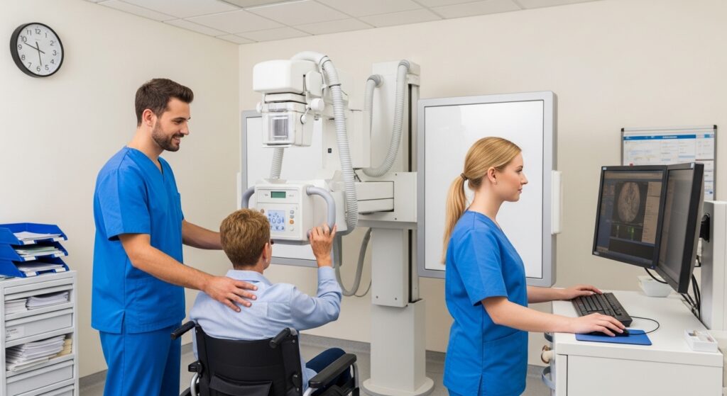
Access X-Ray services as soon as you need them without paying out of pocket. Wosler Diagnostics X-Ray services are covered by all Canadian public health care plans.
If you’re not registered for public healthcare, please contact our clinic before your visit to learn more about X-Ray costs.